
小林 芳郎
マクロファージ-死細胞を取り込んだマクロファージの応答-
はじめに
![]() アポトーシスとは?
アポトーシスとは? ![]() アポトーシス細胞と生きている細胞の違いは何か?
アポトーシス細胞と生きている細胞の違いは何か?
 アポトーシス細胞と生きている細胞の違いは何か?
死んだ細胞は生きている細胞と区別されて、マクロファージなどの細胞によって貪食除去される。
ではアポトーシス細胞と生きている細胞の最も大きな違いは、何だろうか。
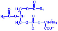
それは細胞膜上のphosphatidylserine (PS)の量(左の図に構造を示す、R1とR2 はアルキル基を示す)である。生きている細胞ではPSはほとんど細胞の内側に局在しているのに対し、アポトーシス細胞ではそれが外側にも出てくる。細胞膜の外側に出てきたPSを認識する分子にはSR-A、 CD14、CD36、MFGE8、Tim4、BAI1 などがある。
もうひとつの違いは糖鎖である。アポトーシスが起こると、糖鎖(ここでは正確には糖鎖を持つタンパク質)の存在状態が変化してクラスターを形成する。この変化をレクチン様の分子が認識する。これにはヌクレオリンが関係するといわれている。その他トロンボスポンジンがアポトーシス細胞を認識するとこれをCD36やαvβ3が認識する。
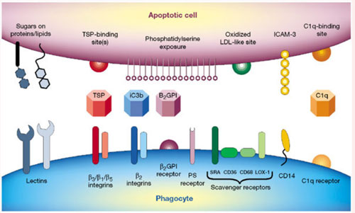
(Nature 407, 784-788, 2000) TSP:トロンボスポンジン
ただしネクローシス細胞でもPSが外側に出てくる場合が報告されているから注意が必要である。
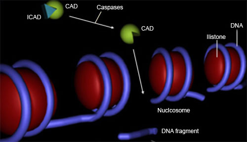
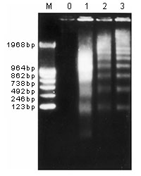
さてアポトーシスが始まると、まずPSが表面に検出されるとともに、細胞サイズが減少し、カスパーゼ(![]() *1)が活性化されて、DNAのラダー様分解(
*1)が活性化されて、DNAのラダー様分解(![]() *2)がおこる。やがて生きた細胞だと透過しない色素(プロピジウムイオダイド、PI、など)で染色されるようになる。ただし細胞質タンパク質(たとえば乳酸脱水素酵素など)の漏出は伴わない。さらに時間が経過すると細胞質タンパク質の漏出が起こる。
*2)がおこる。やがて生きた細胞だと透過しない色素(プロピジウムイオダイド、PI、など)で染色されるようになる。ただし細胞質タンパク質(たとえば乳酸脱水素酵素など)の漏出は伴わない。さらに時間が経過すると細胞質タンパク質の漏出が起こる。
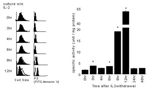
細胞表面のPSをフローサイトメーターで検出するのに蛍光標識アネキシンVがよく用いられる。私たちは、アネキシンV陽性、PI陰性を初期アポトーシス、アネキシンV陽性、PI陽性を後期アポトーシス、細胞質タンパク質の漏出が見られる時期の細胞を2次ネクローシスと呼ぶことにしてきた。下に、IL-2依存性CTLL-2細胞をIL-2非存在下培養して経時的に細胞サイズ、アネキシンVでの染色、カスパーゼ活性を見た結果を示す。9時間目でアネキシンVで染色され始め、カスパーゼ活性が上昇し始めている。なお12時間目ではPIでほとんど染色されない。
  |
しかし研究者によっては、アネキシンV陽性、PI陰性をアポトーシス、アネキシンV陽性、PI陽性を2次的ネクローシスと呼ぶことがある。たとえば組織切片でアポトーシスとネクローシスを区別しようとしている研究者は次のような定義を用いている。
We followed simple yet very useful criteria to define necrosis and apoptosis. Necrosis is defined by the uptake of PI (indicating damage to the cell membrane) and the lack of nuclear condensation and fragmentation. Apoptosis is defined by the lack of PI uptake (indicating the integrity of the cell membrane) and the presence of clear nuclear condensation and/or fragmentation. Secondary necrosis is defined by the presence of nuclear condensation and/or fragmentation along with PI uptake. These are (late) apoptotic cells (and are counted as such) that sustained some terminal damage to the cell membrane before they were cleared.(Am J Physiol Cell Physiol 284, C1309-C1318, 2003)
この研究者は2次的ネクローシスと後期アポトーシスを同義としている(下線部分)。
別の研究者は逆に次のように提案している。
Beyond 18 h in culture, a steadily increasing proportion of senescent neutrophils exhibit a characteristic late apoptotic morphology in which nuclear degradation or so-called evanescence is accompanied by electron microscopic evidence of limited granule fusion, we propose that neutrophils with classical features of apoptosis should be regarded as early apoptotic cells.(J. Immunol. 166, 4743-4750, 2001)
下線部分に示すように、基づく根拠は異なるものの、いわゆるアポトーシスを初期アポトーシスと呼ぶことを提案している。
そこで混乱を避ける目的で、今後はできる限り、初期アポトーシス(ひとによっては単にアポトーシスとよぶ)、あるいは、後期アポトーシス(ひとによっては2次的ネクローシスとよぶ)とできるだけ断ることにする。
大切なことは、正常の個体でアポトーシス細胞を検出するのは一般にとてもむずかしいという点である。これはアポトーシス細胞が現れるや否やすみやかに貪食されているためだと一般に考えられている。たとえば正常なマウス胸腺ではアポトーシス細胞はマクロファージに取り込まれた状態で検出されるという報告がある(Nature 372, 100-103, 1994)。この報告ではアポトーシス細胞をTUNEL法*で検出している。
アポトーシス細胞は本当に現れるや否やすみやかに貪食されているのだろうか。このことを私たちの研究室のSSが蛍光顕微鏡下で微速度撮影を行い調べたところによると何と後期アポトーシス細胞に比べ初期アポトーシス細胞は貪食されにくいという結果が得られた(J Biochem 141, 301-307, 2007)。
さて前置きと準備はこのくらいにして、私たちの研究を紹介しよう。 →私たちの研究
![]()